

NEUROTROPHIC KERATITIS (NK)
NK Is a Degenerative Corneal Disease
Driven by Nerve Impairment1,2
Breakdown of the corneal nerves and surface prevents homeostasis and healing1,2
Click each aspect of the mechanism of disease to learn more
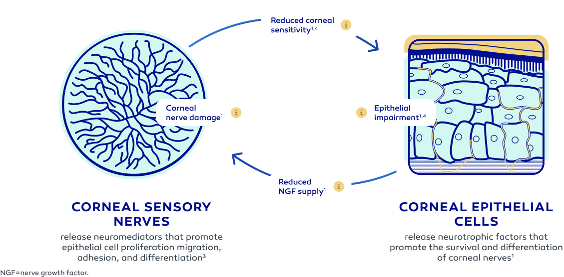
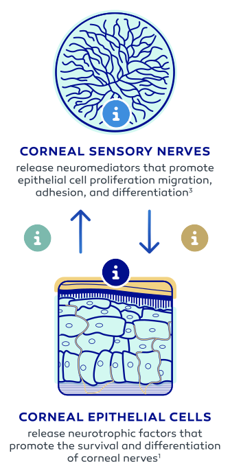
The cascade begins with corneal nerve damage.
Corneal nerve impairment disrupts the ability to sense stimuli that trigger important functions in the cornea.
Reduced ability to sense stimuli impairs normal functions that preserve the health of the ocular surface.
Functions impacted include tear film production and maintenance and the blink reflex, as well as production of factors normally supplied by the nerves to support the health of the corneal epithelium.
When critical functions are impacted, the corneal epithelium cannot maintain its health or repair any damage.
Mitosis of epithelial cells is reduced, preventing healthy cells from replacing dead or damaged ones.
Disruptions to the corneal epithelial cells diminish their ability to supply trophic factors, such as NGF, that support corneal innervation and integrity.
This puts in place a dangerous feedback loop that ultimately impacts the integrity of both the cornea and the corneal nerves.
NGF=nerve growth factor.
NK is driven by corneal nerve impairment that disrupts the normal homeostasis that
maintains the health of both the cornea and the nerves5
Based on preclinical models.1 These images are for illustrative purposes only; they are not an exact representation of structure or function.
Corneal nerve damage can be caused by
common conditions that you see regularly
DRY EYE
DISEASE
of patients with dry eye disease have decreased corneal sensitivity6,*
CORNEAL INSULTS
(IATROGENIC)
of patients with NK had an etiology of prior ocular treatments (eg, surgery, eye drop toxicity)7,†
DIABETES
MELLITUS
of patients diagnosed with NK had a history of diabetes mellitus8,‡
POSTHERPETIC
INFECTION
of patients diagnosed with NK had a history of herpetic keratitis8,‡
Any condition that can cause trigeminal
nerve damage can cause NK.9

CENTRAL NERVOUS SYSTEM (CNS)10

- Neoplasm (acoustic neuroma, meningioma, orbital tumor)
- Aneurysm
- Stroke
- Degenerative CNS disorders
- Postneurosurgical procedures (trigeminal neuralgia, other surgical injury to trigeminal nerve)
SYSTEMIC10

- Diabetes
- Vitamin A deficiency
- Amyloidosis
- Multiple sclerosis
- Sjögren syndrome
- Dry eye disease
- Postherpetic infections
INJURY10

- Ocular nerve lesion, corneal nerve lesion
- Trauma to the ciliary nerve
- Facial trauma
- Chemical and thermal injury
- Ultraviolet light injury
- Radiation
- Prolonged overuse of contact lenses
- Drug-induced (eg, Stevens-Johnson syndrome)
IATROGENIC10

- Chronic use of topical medication
- Surgery (LASIK, keratoplasty, cataract, orbital surgery)
- Panretinal photocoagulation
- Cyclocoagulation
- Benzalkonium chloride exposure
- Abuse of topical anesthetics
GENETICS10

- Goldenhar-Gorlin syndrome
- Moebius syndrome
- Familial corneal hypoesthesia
- Congenital insensitivity to pain with anhidrosis
- Riley-Day syndrome
- Ectodermal dysplasia
OTHER CONDITIONS10

- Vernal keratoconjunctivitis
- Atopic keratoconjunctivitis
- Ocular cicatricial pemphigoid
These conditions should trigger suspicion of NK. This is not an exhaustive list.10
LASIK=laser-assisted in situ keratomileusis.
*Based on a retrospective, single-center chart review conducted on consecutive patients who presented to the eye clinic and underwent corneal sensitivity testing using a noncontact esthesiometer between January 2024 and December 2024. A total of 395 eyes from 198 patients were included for analysis.
†Based on a retrospective, observational, monocenter study, which included patients examined at the Fondation Ophtalmologique Adolphe de Rothschild Hospital, a tertiary referral center, between November 1, 2009, and October 31, 2017. In total, 392 medical records were identified and reviewed for NK.
‡Based on a retrospective observational study using data from the American Academy of Ophthalmology Intelligent Research in Sight® Registry, analyzing 27,483 patients (31,915 eyes) diagnosed with NK between 2013 and 2018.
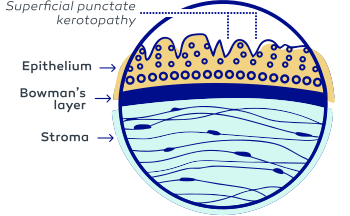
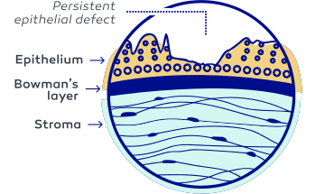
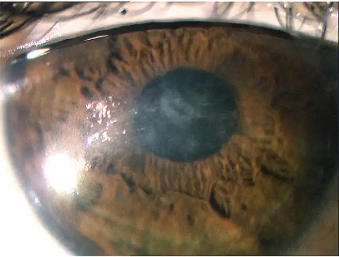
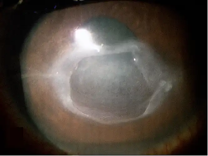
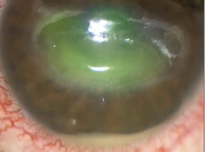
NK can be progressive and occurs in 3 distinct stages2,4
Flip cards to see more
Stage 1
Superficial punctate
keratopathy
Stage 2
Persistent epithelial defect,
stromal swelling
Stage 3
Corneal thinning, ulceration,
perforation, permanent vision loss
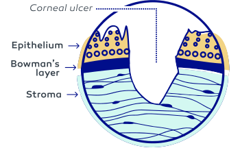
Images sourced from Sacchetti M et al in Clin Ophthal 2014.
Stage 1
Stage 2
Stage 3
Reduced sensation can stop patients from feeling corneal damage,
but there are still symptoms to watch out for4,11,12
Photophobia
(sensitivity and squinting in bright light)

Blurred vision
(may be voiced as trouble reading, using screens, etc)

Reduced
blinking
Dryness
and/or burning

Foreign body
sensation
Indication and Important Safety Information
WARNINGS AND PRECAUTIONS
Use with Contact Lens
Contact lenses should be removed before applying OXERVATE because the presence of a contact lens (either therapeutic or corrective) could theoretically limit the distribution of cenegermin-bkbj onto the area of the corneal lesion. Lenses may be reinserted 15 minutes after administration.
Eye Discomfort
OXERVATE may cause mild to moderate eye discomfort such as eye pain during treatment. The patient should be advised to contact their doctor if a more serious eye reaction occurs.
ADVERSE REACTIONS
In clinical trials, the most common adverse reaction was eye pain following instillation which was reported in approximately 16% of patients. Eye pain may arise as corneal healing occurs. Other adverse reactions occurring in 1% to 10% of OXERVATE patients included corneal deposits, foreign body sensation, ocular hyperemia, ocular inflammation, photophobia, tearing, and headache.
USE IN SPECIFIC POPULATIONS
Pregnancy
There are no data from the use of OXERVATE in pregnant women to inform any drug associated risks.
Lactation
The developmental and health benefits of breastfeeding should be considered, along with the mother’s clinical need for OXERVATE, and any potential adverse effects on the breastfed infant from OXERVATE.
Pediatric Use
The safety and effectiveness of OXERVATE have been established in the pediatric population. Use of OXERVATE in pediatric patients 2 years of age and older is supported by evidence from adequate and well-controlled trials of OXERVATE in adults with additional safety data in children.
INDICATION
OXERVATE® (cenegermin-bkbj) ophthalmic solution 0.002% (20 mcg/mL) is indicated for the treatment of neurotrophic keratitis.
DOSAGE AND ADMINISTRATION
Instill one drop of OXERVATE in the affected eye(s), 6 times a day at 2-hour intervals for eight weeks.
To report ADVERSE REACTIONS, contact Dompé U.S. Inc. at 1-833-366-7387 or FDA at 1-800-FDA-1088 or www.fda.gov/medwatch.
Please see full Prescribing Information for OXERVATE.
References: 1. Mastropasqua L, Massaro-Giordano G, Nubile M, Sacchetti M. Understanding the pathogenesis of neurotrophic keratitis: the role of corneal nerves. J Cell Physiol. 2017;232(4):717-724. 2. Sacchetti M, Lambiase A. Diagnosis and management of neurotrophic keratitis. Clin Ophthalmol. 2014;8:571-579. 3. Müller LJ, Marfurt CF, Kruse F, Tervo TM. Corneal nerves: structure, contents and function. Exp Eye Res. 2003;76(5):521-542. 4. Dua HS, Said DG, Messmer EM, et al. Neurotrophic keratopathy. Prog Retin Eye Res. 2018;66:107-131. 5. Neurotrophic Keratopathy Study Group. Neurotrophic keratopathy: an updated understanding. Ocul Surf. 2023;30:129-138. 6. Stolz M. The prevalence of corneal sensitivity loss in patients with and without dry eye disease. Clin Ophthalmol. 2025;19:1323-1330. 7. Saad S, Abdelmassih Y, Saad R, et al. Neurotrophic keratitis: frequency, etiologies, clinical management and outcomes. Ocul Surf. 2020;18(2):231-236. 8. Bian Y, Ma KK, Hall NE, et al. Neurotrophic keratopathy in the United States: an Intelligent Research in Sight Registry analysis. Ophthalmology. 2022;129(11):1255-1262. 9. Labetoulle M, Baudouin C, Calonge M, et al. Role of corneal nerves in ocular surface homeostasis and disease. Acta Ophthalmol. 2019;97(2):137-145. 10.Vera-Duarte GR, Jimenez-Collado D, Kahuam-López N, et al. Neurotrophic keratopathy: general features and new therapies. Surv Ophthalmol. 2024;69(5):789-804. 11. Ruiz-Lozano RE, Hernandez-Camarena JC, Loya-Garcia D, Merayo-Lloves J, Rodriguez-Garcia A. The molecular basis of neurotrophic keratopathy: diagnostic and therapeutic implications. A review. Ocul Surf. 2021;19:224-240. 12. Murray LT, McCormack J, Grobeiu I, Wiklund I, Kimel M, Van Nooten F. Development of the neurotrophic keratopathy questionnaire: qualitative research. J Patient Rep Outcomes. 2020;4(1):30.
Indication and Important Safety Information
OXERVATE® (cenegermin-bkbj) ophthalmic solution 0.002% (20 mcg/mL) is indicated for the treatment of neurotrophic keratitis.
WARNINGS AND PRECAUTIONS
Use with Contact Lens
Contact lenses should be removed before applying OXERVATE because the presence of a contact lens (either therapeutic or corrective) could theoretically limit the distribution of cenegermin-bkbj onto the area of the corneal lesion. Lenses may be reinserted 15 minutes after administration.
